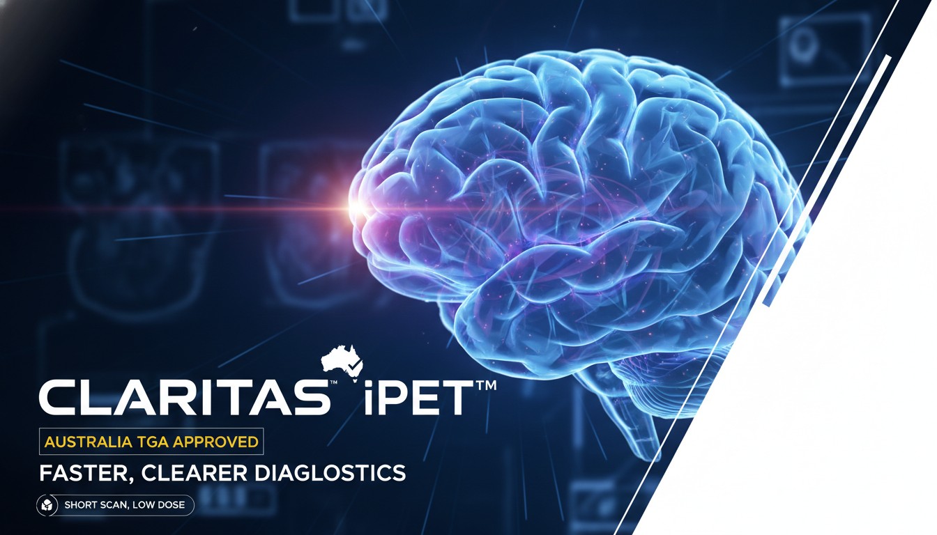
Claritas iPET™, an innovative medical software device from Claritas HealthTech, has secured approval from Australia’s Therapeutic Goods Administration (TGA) to supply its image processing technology. This clearance enables hospitals and imaging centers in Australia to utilize the software for enhancing Positron Emission Tomography (PET) scans, including those combined with CT or MRI. The tool delivers diagnostic-quality images even from scans conducted with significantly shortened durations or reduced radiotracer doses, potentially cutting scan times by up to 80% while maintaining or improving image clarity through noise reduction, boundary sharpening, and super-resolution capabilities. This development expands access to more efficient, patient-friendly nuclear medicine imaging in Australia, building on prior regulatory clearances in the US and other markets.
TGA Approval Marks Key Milestone for Claritas iPET™ in Australian Healthcare
The recent approval by the Therapeutic Goods Administration positions Claritas iPET™ as a Class IIa medical device available for supply across Australia. This regulatory step allows healthcare providers to integrate the software into existing workflows without disrupting standard protocols. The software processes DICOM-format PET images, applying advanced algorithms to improve overall image quality.
Claritas iPET™ focuses on addressing common challenges in PET imaging, where longer scan times and higher isotope doses are traditionally required to achieve sufficient signal-to-noise ratios for accurate diagnosis. By employing sophisticated processing techniques, including non-local means filtering and fusion of functional PET data with anatomical details from CT or MRI overlays, the software enhances visibility of structures and lesions.
Key technical features include:
Noise reduction — Effectively suppresses background noise in low-signal scans, making subtle abnormalities more detectable.
Boundary sharpening — Improves delineation of organ and lesion edges, aiding precise localization in oncology, cardiology, and neurology applications.
Super-resolution enhancement — Increases effective resolution beyond the original acquisition, allowing clinicians to extract more diagnostic information from the same data.
User-adjustable parameters — Radiologists and nuclear medicine physicians can fine-tune the degree of enhancement to suit specific clinical needs or preferences.
One of the most impactful benefits is the potential for substantial reductions in scan duration. In practice, facilities using similar enhanced processing have reported shortening PET acquisitions significantly while preserving diagnostic confidence. This translates to greater patient comfort—particularly important for those with claustrophobia, pain, or movement issues—and higher throughput in busy imaging departments. Reduced scan times also mean lower exposure to radiation for patients when isotope doses are decreased, aligning with ALARA (As Low As Reasonably Achievable) principles.
The software remains vendor-agnostic, compatible with major PET, PET-CT, and PET-MRI systems from various manufacturers. Implementation requires no hardware modifications or changes to acquisition protocols, making adoption straightforward for radiology and nuclear medicine groups.
In oncology, where PET scans play a central role in staging, treatment response assessment, and recurrence detection, clearer images from lower-dose or faster scans can support earlier interventions and more accurate monitoring. For cardiac viability studies or neurological applications like dementia evaluation, the enhanced contrast and reduced noise contribute to better quantification of metabolic activity.
This TGA clearance follows a series of international milestones for the platform, including 510(k) clearance from the US FDA several years ago, along with approvals in markets such as India and Brazil. The expansion into Australia reflects growing global recognition of AI-driven and algorithm-based image enhancement tools in nuclear medicine. As demand for PET imaging continues to rise—driven by an aging population and increasing cancer incidence—the ability to optimize scan efficiency becomes increasingly valuable.
Healthcare systems in Australia, like those elsewhere, face pressures to manage rising caseloads while controlling costs and minimizing patient burden. Claritas iPET™ offers a software-only solution that can help address these issues by enabling more scans per day and reducing the need for repeat imaging due to suboptimal quality.
Clinicians gain tools that streamline interpretation, with improved image quality facilitating confidence in findings even under challenging acquisition conditions. The platform’s focus on maintaining diagnostic equivalence to standard scans ensures it meets rigorous clinical standards.
As nuclear medicine evolves with hybrid modalities and personalized approaches, technologies like Claritas iPET™ support the shift toward more efficient, lower-risk imaging protocols. Australian providers now have access to this capability, potentially transforming how PET studies are performed and interpreted in clinical settings nationwide.
Disclaimer: This is a news report on regulatory developments and medical technology advancements. It is for informational purposes only and not investment, medical, or regulatory advice.